Highlights
Highlights
Brain development: Made from girders
- 2010/03/25
The girder protein has a critical role in multiple stages of neuron development
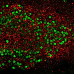
Fig. 1: In mutant mice lacking the Girdin protein, immature neurons migrate too far into the dentate gyrus region of the hippocampus (top).
During neural development in mammals, huge numbers of neurons are generated at the lining of the major brain cavities, called the cerebral ventricles. Newborn cells migrate from their birthplace to their final destination, before extending their axons and dendrites to form connections with other cells. A new study involving researchers from the Nagoya University GCOE for Molecular Medicine1 has now shown that a protein called girdin plays multiple roles in postnatal development of the hippocampus, a part of the brain known to be critical for memory formation. Girdin -- short for 'girders of actin filaments' -- is known to bind to and remodel the actin cytoskeleton in neurons, and also to be involved in cell movements. The research team, led by Atsushi Enomoto from the Nagoya University Graduate School of Medicine, used antibody staining to show that girdin is expressed abundantly in the hippocampus of mice both before and after they are born. Girdin interacts with a protein called disrupted-in-schizophrenia 1 (DISC1), which is implicated in numerous developmental processes including the migration and differentiation of hippocampal neurons. The researchers found that girdin is initially expressed in the cell body of hippocampal neurons, but then localizes to growing axons and accumulates in the extending tip. This distribution is perturbed by inhibiting DISC1 expression, suggesting that DISC1 regulates girdin localization. What's more, when the researchers reduced girdin levels by RNA interference, they saw impairments in axon extension and a reduction in the number of axons and dendrites. To further investigate the role of girdin, the researchers generated mutant mice lacking the girdin gene. The mice exhibited abnormal cellular architecture in the hippocampus: the dentate gyrus region exhibited a slight increase in cell numbers (Fig. 1), whereas other regions had too many layers because of prolonged cell migration or contained cells that were packed too loosely. These defects were also observed in neurons expressing a shortened form of girdin that does not interact with DISC1. An examination of the mutant mice revealed that the defects are at least partly due to impaired axon extension in dentate gyrus cells. The researchers also observed a decrease in the volume of the olfactory bulb, the brain structure that provides a sense of smell. The girdin-DISC1 interaction is therefore critical for the generation and migration of hippocampal and olfactory neurons, as well as the proper formation of axons, dendrites and blood vessels in the dentate gyrus. Because DISC1 is associated with susceptibility to schizophrenia and bipolar disorder, the findings also support the hypothesis that psychiatric disorders occur as a result of abnormal developmental processes.
Affiliated Researchers
The Nagoya University affiliated researchers mentioned in this highlight are from the Integrated Functional Molecular Medicine for Neuronal and Neoplastic Disorders GCOE program of the Department of Pathology and the Graduate School of Medicine.
Reference
- Enomoto, A., Asai, N., Namba, T., Wang, T., Kato, T., Tanaka, M., Tatsumi, H., Taya, S., Tsuboi, D., Kuroda, K. et al. Roles of disrupted-in-schizophrenia 1-interacting protein girdin in postnatal development of the dentate gyrus. Neuron 63, 774-787 (2009). | article