Highlights
Highlights
Bio-imaging: Fat cells get the quantum treatment
- 2010/04/29
A low-toxicity polypeptide can be used to transfer fluorescent nanoprobes into stem cells for imaging
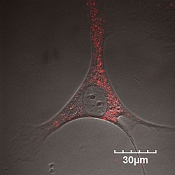
Fig. 1: Confocal laser scanning micrograph of adipose tissue-derived stem cells labeled with quantum dots (red).
c 2010 M. Watanabe
Stem cells are expected to have a major role in regenerative medicine because they can differentiate into various cell types. Adipose tissue-derived stem cells (ASCs), which are abundant and easily harvested from body fat, offer a promising alternative to bone marrow-derived stem cells.
Now, a collaborative team led by Shuji Hayashi and Yoshinobu Baba from the FIRST Research Center for Innovative Nanobiodevices has developed a labeling method that delivers quantum dots -- fluorescent semiconductor nanocrystals -- into ASCs using a polypeptide1.
Unlike conventional dyes, quantum dots are attractive cellular probes with exceptional fluorescence and photostability. They have been shown to spontaneously enter certain cells, but they need carriers to mediate their transfer into ASCs. Carriers like liposomes -- tiny fatty acid pockets that can be filled with molecules -- could achieve this task in a few hours. However, they are generally toxic to cells, even at low concentrations.
To address this problem, Baba and his co-workers chose a low-toxicity, high-efficiency cell-penetrating peptide (R8) as a carrier. They mixed the R8 peptides with negatively charged quantum dots made of cadmium-based semiconductor nanocrystals coated with a zinc-based semiconductor layer. To transfer the quantum dots into the cells, they simply incubated the resulting R8-quantum dot complex with ASCs in a cell transduction medium at 37 °C.
"We selected the R8 peptide because of its similarity to an HIV peptide sequence which is efficient at transfecting certain molecules into cells," says Baba. "Our approach is simpler, easier to handle, less toxic and more efficient than existing methods."
The researchers found that the R8-quantum dot complex labeled more than 80% of the cells in one hour, and at substantially reduced toxicity (Fig. 1). To ensure that the labeling did not affect the ability of ASCs to differentiate into any cell type, they cultured the labeled cells in two separate differentiation media. They discovered that the labeled ASCs contained fat in a fatty tissue-promoting medium and adopted a bone cell-like structure in a bone cell-stimulating medium.
Finally, the team injected labeled ASCs subcutaneously into mouse backs and intravenously into mouse tail veins, and detected their fluorescence for up to seven days after transplantation.
The researchers believe that this imaging technique will provide crucial information on the behavior, localization and condition of transplanted stem cells. "We hope to apply this technology to various stem-cell transplantation therapies, such as islet transplantation," says Baba.
Affiliated Researchers
The Nagoya University affiliated researchers mentioned in this highlight are from the FIRST Research Center for Innovative Nanobiodevices.
Reference
- Yukawa, H.,* Kagami, Y., Watanabe, M., Oishi, K., Miyamoto, Y., Okamoto, Y., Tokeshi, M., Kaji, N., Noguchi, H., Ono, K., Sawada, M., Baba, Y., Hamajima, N., Hayashi, S. Quantum dots labeling using octa-arginine peptides for imaging of adipose tissue-derived stem cells. Biomaterials 31, 4094-4103 (2010). article