Highlights
Highlights
Magnetic materials: A clearer picture
- 2010/06/29
An ultrabright source of spin-polarized electrons can be used to observe the growth of magnetic materials
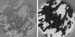
Fig. 1: Snapshots of the growth of cobalt on tungsten by real-time SPLEEM. The development of magnetization (light and dark areas) was observed over just 0.4 seconds as the cobalt increased from 5 monolayers (left) to 7 monolayers (right) in thickness. Images show areas 6 micrometers across.
From Ref. 1. Reproduced with permission. c 2010 The Japan Society of Applied Physics
Advances in electronics and computer technology are becoming increasingly dependent on our understanding of magnetic materials. The growth of these materials can now be imaged in real time thanks to the work of a team of researchers in Japan led by scientists from Nagoya University1.
The researchers took a well-established technique, spin-polarized low-energy electron microscopy (SPLEEM), and refined it to be able to take snapshots of a magnetic material in just a fraction of a second. SPLEEM probes the properties of a material by bombarding a sample with a stream of spin-polarized electrons, allowing the magnetic properties of a material to be measured with a resolution of just 10 nanometers. However, a single SPLEEM image can take up to 10 seconds to acquire. The key to extending the technique to real-time imaging was the development of a brighter electron beam with higher spin purity.
In conventional SPLEEM, the electron beam is produced by focusing an electron beam onto an electron-emitting cathode surface. The laser excites the emission of electrons back in the direction of the laser, which means the laser itself must be set a relatively long distance from the cathode, limiting how small and intense the laser spot on the cathode can be.
To enhance the intensity of electron production, the research team created their electron source on a transparent base and instead shone the laser from behind. "The challenge has been creating a transparent cathode with the same quality as an opaque substrate," explains Yoshikazu Takeda, one of the Nagoya University researchers on the team. With this new design, the scientists were able to focus the laser to a spot as small as 1.3 micrometers in diameter. "In this way, the brightness of the beam is increased by four orders of magnitude," says Takeda.
The researchers built their transparent cathode from alternating layers of gallium arsenide and gallium arsenide phosphide. By carefully controlling the strain that builds up in these layers, they could increase the purity of electrons emitted with the correct spin polarization to as high as 90%.
These two design elements result in a 'spin gun' that is perfectly suited to real-time imaging of magnetic materials. A SPLEEM system incorporating this electron source was successfully used to observe the growth of magnetic domains in layers of cobalt as they were deposited on a tungsten substrate (Fig. 1), allowing the researchers to observe how some domains vanished as the cobalt became thicker.
More detailed knowledge of magnetic materials could aid researchers in a number of different areas. "One example could be hard disks for memory," suggests Takeda. "We can learn how small a domain can be made without losing the magnetized, or memorized, region."
Affiliated Researchers
The Nagoya University affiliated researchers mentioned in this highlight are from the Department of Crystalline Materials Science, Graduate School of Engineering, and the Department of Particle and Astrophysical Science, Graduate School of Science.
Reference
Suzuki, M., Hashimoto, M., Yasue, T., Koshikawa, T., Nakagawa, Y., Konomi, T., Mano, A., Yamamoto, N., Kuwahara, M., Yamamoto, M. et al., Real time magnetic imaging by spin-polarized low energy electron microscopy with highly spin-polarized and high brightness electron gun. Applied Physics Express 3, 026601 (2010). | article