Highlights
Highlights
Cell engineering: Patterning behavior
- 2010/10/28
The fabrication of microscale biomolecular arrays just got simpler thanks to new design rules for controlling cell adhesion
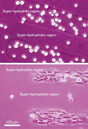
Fig. 1: Microscope images of cell cultures on a micropatterned surface after one hour (top) and one day (bottom). Cells initially preferentially cluster in superhydrophilic regions, but after one day, nearly all cells had spontaneously migrated to superhydrophilic zones.
c 2010 ACS
The process of arranging living cells in ordered arrays on micropatterned polymer surfaces is essential for technologies such as drug screening and artificial tissue growth. Unfortunately, current methods of attaching cells to surfaces can sometimes produce anomalous and poorly resolved cell patterns. Nagahiro Saito from the EcoTopia Science Institute at Nagoya University1 and co-workers have now established a set of empirical design rules that makes the production of two-dimensional cellular microarrays easier than ever.
One of the best ways to position cells in surface arrays is to create alternating water-repelling and -attracting regions on a substrate. Then, by culturing a watery suspension of cells over the surface, the biomolecules spontaneously attach to the 'superhydrophilic' zones while remaining clear of 'superhydrophobic' areas. Occasionally, however, cells adhere in the water-repelling regions. Saito and his colleagues set out to resolve this dilemma by investigating the effects of pattern topography and geometry in these systems.
The team produced model microcellular arrays by first depositing a superhydrophobic silicon-hydrocarbon film onto a glass slide. They then created two-dimensional arrays of circular superhydrophilic regions in the film by using ultraviolet light to convert the exposed hydrocarbons into water-attracting hydroxyl groups.
The researchers established a mouse fibroblast cell culture on the patterned surface and found that after one hour, the cells selectively clustered in the superhydrophilic regions (Fig. 1). Within 24 hours, even those cells that did not initially stick to the array spontaneously migrated to the binding zones. However, this form of cell-to-cell recognition can lead to problems: the team saw that if distances between superhydrophilic regions were less than 250 micrometers, the cell cultures reached beyond their initial patterns and coalesced together.
After three days of incubation on the micropatterned arrays, a surprise awaited the research team: the cells adhered over the entire surface, including the superhydrophobic areas. "Up until 72 hours, we only saw cell death in these areas," notes Saito. "Afterwards, however, it was as if the cell's graveyard changed into their home."
This unexpected behavior was shown to be caused by surface roughness. The bumpy topography of the superhydrophobic film provided enough surface area to establish a collagen-like matrix for nucleating cell growth, given enough incubation time.
With these design guidelines in place, the researchers are set to take on their next challenge: organizing stem cells into micropatterned arrays. "If we can control stem cells, it will help us to conduct tissue engineering and to develop drug screens for cancer," says Saito.
Affiliated Researchers
The Nagoya University affiliated authors mentioned in this highlight are from the EcoTopia Science Institute and the Department of Materials Science and Engineering.
Reference
Ishizaki, T., Saito, N. & Takai, O. Correlation of cell adhesive behaviors on superhydrophobic, superhydrophilic, and micropatterned superhydrophobic/superhydrophilic surfaces to their surface chemistry. Langmuir 26, 8147-8154 (2010). | article