Highlights
Highlights
Discovery of a new memory mechanism―expecting infinite practical use
- Read in Japanese
- 2016/02/17
- Graduate School of Science
- Mr. Kyogo Kobayashi (a researcher)
- Prof. Ikue Mori
“Nothing is ruined.” Challenging spirits of young researchers can open the way to unveil a new theory.
"Humans conduct themselves based on their emotions, such as pleasure and unpleasure."
Prof. Ikue Mori and her research group at the Graduate School of Science at Nagoya University seek to solve challenging questions on neurological mechanisms. The researchers used the nematode Caenorhabditis elegans as a model organism in their experiments because many of the functional molecules involved in the C. elegans nervous systems are known to have the same activity as their counterparts in humans.
Their research was performed in three steps: first, observe the behavior of C. elegans related to its thermotaxis; second, the associated neuron is identified; and third, the specific genes or molecules involved are characterized. In particular, they concentrated on the function of the nervous system as it relates to memory mechanisms.
Despite the current compelling theory on the mechanism of animal memory (i.e., synaptic plasticity between nervous cells), the researcher’s study showed a new memory function of synapse-independent cells at the molecular level. Thus, this new discovery could rewrite the widely accepted theory of memory.
In basic research, the importance of achievements is immeasurable for the future development. This study is also expected to contribute to the definitive understanding of memory mechanisms and help advance the treatment of neurological diseases.
*******
—Mori Laboratory, the research lab of “thermotaxis behavior of C. elegans”
“Even these tiny creatures can change their behavior depending on rewards.”
The nematode C. elegans has a transparent body about 1 mm in length and usually lives in temperate soil environments.
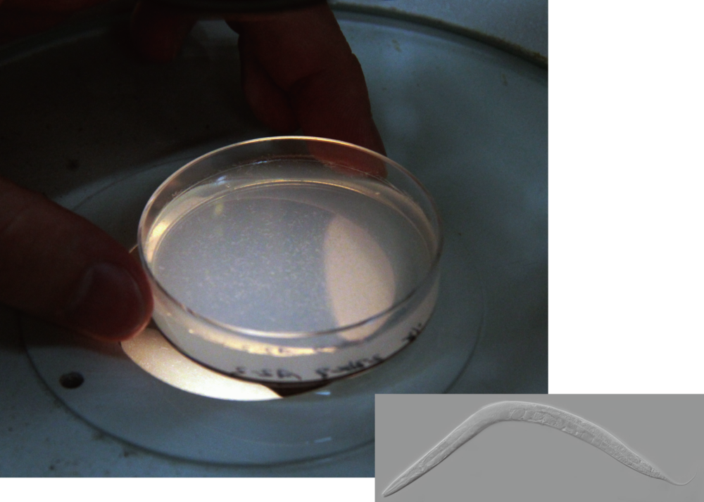 The nematode C.elegans on culture medium (left), and its single form (right bottom).
Although the C. elegans body form is tremendously different from that of humans, they have in common pleasure/unpleasure emotions controlled by monoamine neurotransmitters such as serotonin. C. elegans is a model organism that can be utilized in research on human nerve cells to overcome the difficulty in using humans for research.
The human brain has more than 100 billion nerve cells. There are only 302 neurons in C. elegans, and all of them have been identified and individually named with three letters of the alphabet.
For example, AFD is a thermosensory neuron identified by Prof. Mori. It is located upstream of the neural circuit controlling thermosensory behavior and is related to actions caused by temperature dependence or thermotaxis (Figure 1). (Mori and Ohshima, Nature (1995) 376, 344-8)
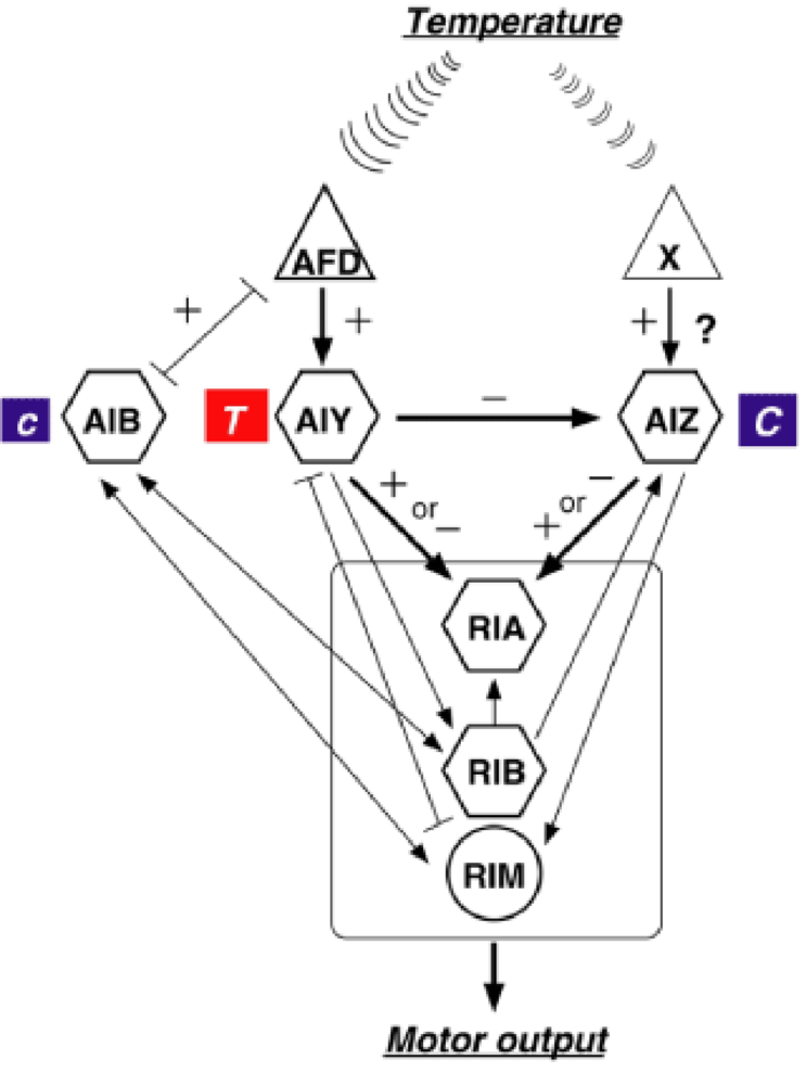
Figure 1. Neural circuits and behavioral traits generating thermotaxis (souce : Research 1, Mori Laboratory HP accessed on January 28, 2016)
—The original meaning of “temperature” dependence, revealed by visualization
To visualize C. elegans behavioral changes caused by temperature, Prof. Mori and her research group introduced a new technique called calcium imaging. They found out that the AFD neuron intracellular calcium concentration increases in response to a temperature changing.
In 2004, Dr. Kotaro Kimura, a postdoctoral researcher in the same research group at that time, measured the level of calcium ion concentration inside the AFD neuron by monitoring the intensity of fluorescence from a sensor molecule that reacted to changes in calcium concentration. This research showed that the calcium level of AFD increases around the cultivation temperature (Figure 2). (Kimura, et al. Current Biology (2004) 14, 1291-5)
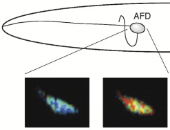
Figure 2. For the physiological analysis of thermotaxis by measuring the level of calcium ion concentration inside the AFD neuron (<blue> before activation/low calcium ion concentration; <red> after activation/high calcium ion concentration) (souce : Research 1, Mori Laboratory HP accessed on January 28, 2016)
The research group then determined the original meaning of temperature dependence, that is, thermotaxis action in the AFD neuron.
As C. elegans is active between 15°C and 25°C, organisms cultivated at 15°C or at 25°C moved toward a lower temperature (~15°C) or a higher temperature (~25°C), respectively. This finding showed the high importance of calcium ion concentration inside the AFD neuron. Thus, the AFD neuron not only detects temperature, but it also depends on the cultivation temperature for some of its functionality.
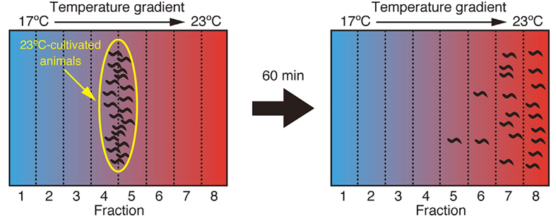 Figure 3. The nematode C. elegans cultivated at 23℃ moved to 23℃ after 60 mins.
“We were surprised. AFD exhibits a memory function as well as detects the ambient temperature.”
Prof. Mori and her research group doubted the commonly accepted theory on the mechanism of memory because of their observations that the neuronal activity of the AFD neuron depended on the cultivated temperature.
—“However, how can we verify this observation experimentally?” Prof. Mori asked. If the AFD neuron can save specific memorized information, it would remain as a memory even if some parts in the neural circuit are disconnected.
—Required for a new experimental system
“I want to solve the question of memory mechanism using C. elegans.”
Mr. Kyogo Kobayashi, a researcher in the same research group, was especially interested in the work of the Mori Laboratory when he was a senior student looking for a laboratory in which to perform his graduation work.
After joining the laboratory, he started research with the theme of an analysis of functioning molecules within AFD neurons. Despite his extreme initial motivation, he found it difficult to establish the next research step toward that end.
Prof. Mori then encouraged Mr. Kobayashi by suggesting another research theme to him. To reveal the thermotaxis behavior caused by AFD neural activity as described above, she explained to him that a new experimental system capable of evaluating the AFD neuron before memory system development had to be established.
Mr. Kobayashi was inspired by the idea and decided to work in a master’s course on this new challenge. He started by first making a primary culture system to isolate a neural cell from the fertile ovum of C. elegans related to the thermotaxis behavior.
At that time, however, utilizing a C. elegans primary culture system for experimentation was not common, and Mr. Kobayashi himself did not possess the basic skills needed to maintain the cultivated cells. Although he followed published methods of establishing a primary culture system, the technique was not that easy, and his challenges continued for more than a year.
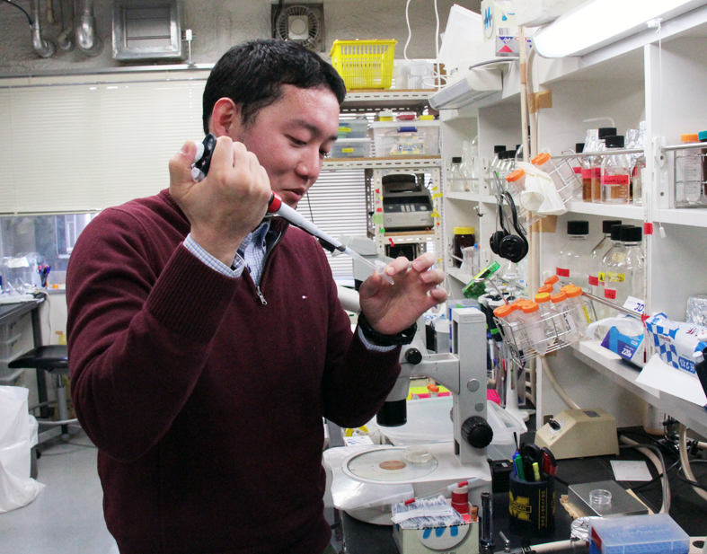 Mr. Kobayashi in the lab
“Still, by doing so, I started to understand that the composition of the culture medium was important.”
Learning through trial and error, Mr. Kobayashi found a hint in an electrophysiological study of C. elegans. In electrophysiology, one of the experimental methods is to insert the tip of the electrode into a single cell to observe and record the electrical activity of that cell. In this way, cells are exposed outside the body. To keep them alive, artificial extracellular fluid is required.
According to the composition of the extracellular fluid, Mr. Kobayashi then finally created a culture medium. In fact, establishing a primary culture system for AFD neurons was a significant step toward success.
Utilizing the primary culture system, he experimented with calcium imaging and noted that the calcium level in isolated AFD neurons increased depending on the previous culture temperature. That is, memory formation in an AFD neuron was surmised to be independent and to not require interaction with other neurons (Figure 4).
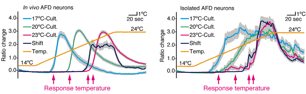 Figure 4. Calcium imaging of in vivo AFD cultivated (Cult.) (left) and isolated AFD neurons cultured (right). Memory formation in an AFD neuron was surmised to be independent and to not require interaction with other neurons.
—For identification of the genes or molecules
“Further development of research and applied use with humans requires a definitive understanding of occurrences at a molecular level.”
Mr. Kobayashi performed continuous experiments using different gene mutants to define the mechanism of memory formation by isolated AFD neurons at molecular level. Checking the common memory-related genes in both of C. elegans and humans, he specifically found the serious anomaly, cmk-1 mutants, that lacked the functionality of C. elegans.
This cmk-1 gene encodes the CMK-1 protein (calcium-calmodulin-dependent protein kinase I/IV, CaMK I/IV), one of key molecules that establish memory. Humans have two CaMKs (CaMK I and CAMK IV) that are homologous to the C. elegans CMK-1 and can act as enzymes to phosphorylate brain proteins. However, their functions and interacting molecules that target phosphorylation are largely unknown.
The research group asked a collaborative research team led by Prof. Kozo Kaibuchi at the Graduate School of Medicine at Nagoya University for a phosphoproteomic screen test to look for biochemical substrates of CMK-1. The screen identified 38 proteins from among all of the proteins expressed by C. elegans as molecules phosphorylated by human CaMK I.
“My graduation work at that time turned out to be useful here!”
From these 38 proteins, Mr. Kobayashi immediately found out that, in particular, the lack of Raf kinase caused defects in AFD neuron memory. The data related to Raf kinase were actually obtained when he was a senior student performing his graduate research work.
Because of this finding, his subsequent studies showed rapid progress and led to a new understanding of the neural cell function; that is, Raf kinase pathway (CaMK I/IV–Raf–MEK–ERK–MED23) regulation of the single-cell memory system active in C. elegans and humans was unveiled (Figure 5).
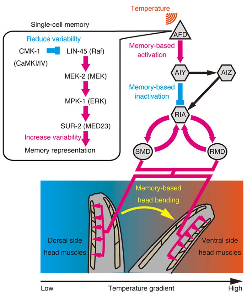 Figure 5. Summary of single-cell memory system in the nematode C. elegans.
“The data were so meaningful then!”
Prof. Mori was also pleased to hear when Mr. Kobayashi had obtained these results. Some people may think that basic research can be drudgery; however, every aspect of the research process is meaningful. “Furthermore, practical applications such as molecular targeted drugs or applications in pharmaceutical development can be expected,” says Mr. Kobayashi. The potential utility of building upon these basic research results is infinite.
*******
“What we are doing in research is fully transparent in our laboratory.”
Prof. Mori guides students and young researchers through frank discussions that help decide the purpose and methodology of research. In these discussions, the results obtained are also critically evaluated from various perspectives. Once students have obtained a doctorate title, they become professionals. Prof. Mori is the mentor who can develop young researchers using this policy.
“I enjoy the discussion.”
As Mr. Kobayashi says, mentors help young researchers not only to cope with research difficulties but also to provide opportunities that ultimately allow them to achieve useful research results.
Never lower your standards as a researcher
—thus, you can only grow up.
(Ayako Umemura)
Researchers featured in this article
Dr. Ikue Mori【Professor, Graduate School of Science, Nagoya University】
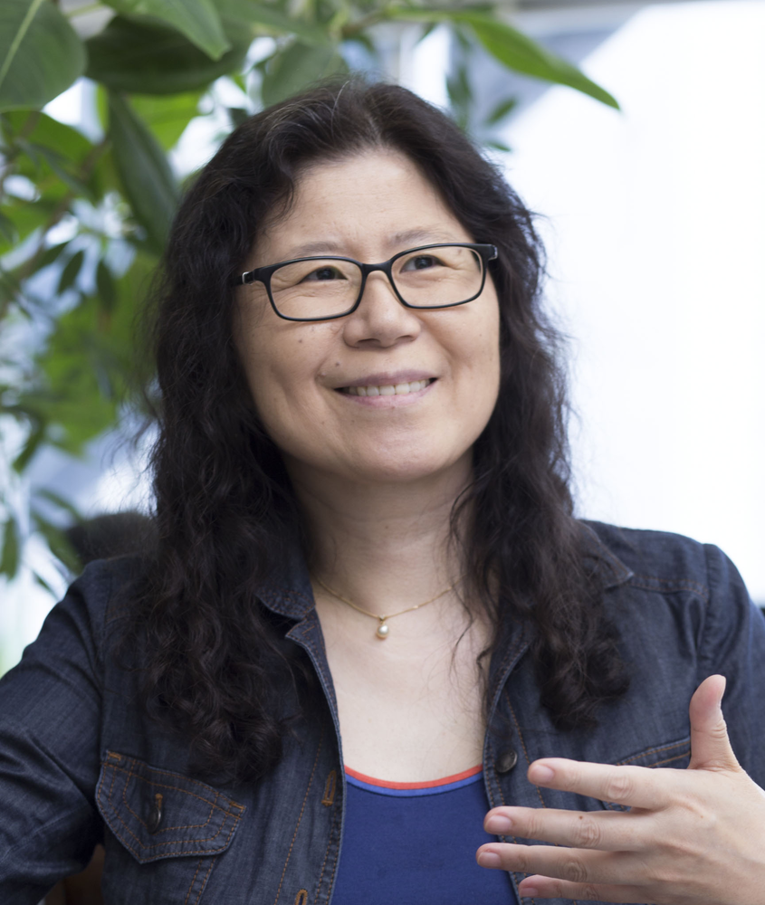 Dr. Mori graduated from the Department of Biology, Faculty of Science, Ochanomizu University in 1980. In 1982, she studied as a visiting graduate student at the University of Sussex with the support of MESSC, Japan, and then obtained a master’s title from Ochanomizu University in 1983. Subsequently, through research conducted as a visiting graduate student at the National Cancer Center Research Institute, she obtained a Ph.D. from Washington University in 1988. Between 1989 and 1998, she worked as an assistant professor at Kyushu University. In 1998, she became a principle investigator as an associate professor at Nagoya University, and since 2004, she has held the title of professor at Nagoya University.
***
“All graduate school experiences are the fundamentals of my research activities,” says Dr. Mori. During her doctorate studies in the United States, in a simulating research atmosphere, she often encountered great researchers who visited her laboratory and some of them became Novel laureates later. “It is the standard of my research life,” she stated, so she teaches young researchers based on her experiences.
In the United States, postdoctoral researchers have been producing a large volume of research results over a short period—Dr. Mori shows high spirits about that. I am looking forward to hearing of future achievements from the Mori Laboratory (by AU)
Mr. Kyogo Kobayashi【Researcher, Graduate School of Science, Nagoya University】

Mr. Kobayashi graduated from the School of Science, Nagoya University in 2009 and then completed his master’s course in the Graduate School of Science at Nagoya University in 2011. Between April 2011 and March 2014, Mr. Kobayashi pursued his research during a doctoral course at the Graduate School of Science at Nagoya University working as a fellow of the Japan Society for the Promotion of Science (DC1). After completing the doctoral course, he started working as a researcher.
***
“Everything is meaningful,” Mr. Kobayashi learned through his research. Like him, research life provides just as a good of a life lesson, which young researchers are strongly and freely learning. Experiences gained here will expand their abilities and widely influence society.
I heard one top researcher says the complimentary statement that “Nagoya University’s young researchers are doing very well.” I would like to cheer all, with this encouragement (by AU)
Links
- Mori Laboratory HP http://elegans.bio.nagoya-u.ac.jp/~lab/index.html
- Graduate School of Science HP http://www.sci.nagoya-u.ac.jp/en/index.html
- Link to this article (from a search-result)
http://www.sciencedirect.com/science/article/pii/S2211124715014205
Kyogo Kobayashi, Shunji Nakano, Mutsuki Amano, Daisuke Tsuboi, Tomoki Nishioka, Shingo Ikeda, Genta Yokoyama, Kozo Kaibuchi, and Ikue Mori.
Single-Cell Memory Regulates a Neural Circuit for Sensory Behavior.
Cell Reports. 14: 11 (2016).
(First published on December 24, 2015; doi: 10.1016/j.celrep.2015.11.064)
- Link to related article1 (from a search-result)
http://www.nature.com/nature/journal/v376/n6538/abs/376344a0.html
Ikue Mori and Yasumi Ohshima.
Neural regulation of thermotaxis in Caenorhabditis elegans.
Nature. 376: 344 (1995).
(First published on July 27, 1995; doi: 10.1038/376344a0)
- Link to related article2 (from a search-result)
http://www.sciencedirect.com/science/article/pii/S0960982204004737
Koutarou D Kimura, Atsushi Miyawaki, Kunihiro Matsumoto, and Ikue Mori.
The C. elegans Thermosensory Neuron AFD Responds to Warming.
Current Biology. 14: 1291 (2004).
(First published on July 26, 2004; doi: 10.1016/j.cub.2004.06.060)