Highlights
Highlights
To increase the reliability of green fluorescent protein (GFP) use
- Read in Japanese
- 2015/01/29
- Graduate School of Bioagricultural Sciences
- Dr. Shoji Segami
- Prof. Masayoshi Maeshima
For the development of science and technology, fundamental research is significant. If researchers use inappropriate markers in a research field of molecular cell biology, the observations will lead to wrong conclusions that we have even never imagined.
"That's something strange, isn't it?"
By doubting a certain phenomenon, Dr. Shoji Segami and his co-workers at the Graduate School of Bioagricultural Sciences, Nagoya University were led to a discovery.
Green fluorescent protein (GFP), a remarkable brightly glowing protein first observed in the jellyfish Aequorea Victoria by Dr. Osamu Shimomura [a Nobel laureate in chemistry (2008)], is widely used as a biological marker to visualize molecules in bioscience. Protein molecules cannot be seen with our naked eyes. By inserting GFP into the target proteins and transferring these genes into cells, GFP enables us to detect the intracellular localization of the proteins using a fluorescence microscope or a confocal laser scanning microscope.
There have been a large amount of data reported by researchers; however, Dr. Segami and his colleagues have not been just trusting them but also been readily checking and studying the phenomena by themselves. In this manner, they have found, besides the ability of GFP to visualize molecules, GFP has the ability to stick to each organelle in a cell.
*******
In the Graduate School of Bioagricultural Sciences, Nagoya University, Prof. Masayoshi Maeshima and his research group focus on molecules that play an active role in the cells that support plant life. Targeting these functional molecules, they aim to increase the production and the expansion of cultivation areas by understanding the mechanism underlying mass transport and energy conversion in biological membranes.
"When focusing on molecules, it is essential to carefully observe their functions in relation to their shape. In this manner, we can determine the physiological phenomena and can control them."
The research attitude of Prof. Maeshima is also seen in his discovery of unknown molecules in vacuole membranes (Figure 1), which was "the first step of independent research" for him (Maeshima and Yoshida, JBC (1989) 264: 20068-73).
The molecule discovered and identified by Prof. Maeshima was the H+-translocating inorganic pyrophosphatase (H+-PPase). H+-PPase is the protein that facilitates the active transport of hydrogen ions (protons) into vacuoles using the energy obtained from the hydrolysis of pyrophosphate, making the vacuoles acidic. Therefore, as an example, the system maintains the essential environment for salt-tolerant plants.
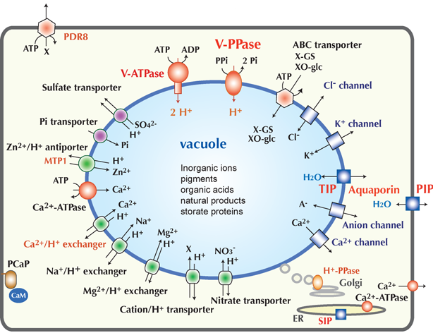
Figure 1. The schematic diagram of plant membrane transport system. Words in red are the research subjects persued by Prof. Maeshima and his research group. (Figure: by courtesy of Prof. Maeshima)
"You may find a discovery on the way to your purpose. Develop further your discovery as your own research."
Under the supervision of Prof. Maeshima, Dr. Segami targeted H+-PPase on the vacuolar membranes and attempted to reveal how vacuoles change their shape under the growth of plants and to also clarify its role.
First, Dr. Segami challenged to associate GFP to H+-PPase to visualize its location and shape.
Because the linkage of GFP to the end of the protein as in the general method did not glow well, GFP was fused to the interior of the protein. So as not to impair the function of H+-PPase, he devised a linkage with soft peptides that can freely change its structure (Figure 2).
 Figure 2. The schematic diagram of GFPs fused to the interior of the protein. (Figure: by courtesy of Dr. Segami)
It was therefore possible to visualize molecules without losing the function under a confocal laser scanning microscope.
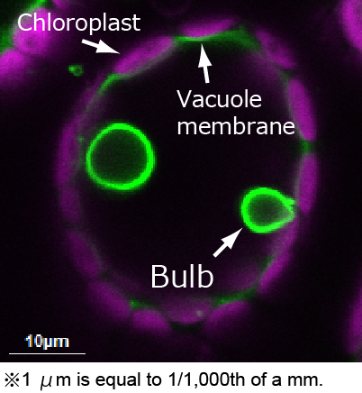
As seen in Figure 3, the structure appeared to be bulb-shaped (similar to light bulbs) labeled by an extremely high intensity of fluorescence.
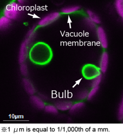 Figure 3. The image of mesophill cell observed by a confocal laser scanning microscope. Green: GFP, magenta: chloroplast. (Figure: by courtesy of Dr. Segami)
In several studies, bulb-like structures have been also reported to be a part of vacuole structures with an unknown function, and because it has been seen in wild-type cells, the bulb-shape has been considered to occur naturally.
By analyzing the various variants in detail, Dr. Segami and his colleagues realized that those bulbs are unnatural artificial products that increase with the amount of GFP molecules. Furthermore, although the force of dimerization of GFPs is generally considered very weak, Dr. Segami suspected that it can act as a glue to stick membranes to each other.
Following his thought, Dr. Segami prepared a monomeric GFP by replacing the location (alanine, 260th residue) suspected to dimerize with lysine (an amino acid with a long positive side chain), and then used it.
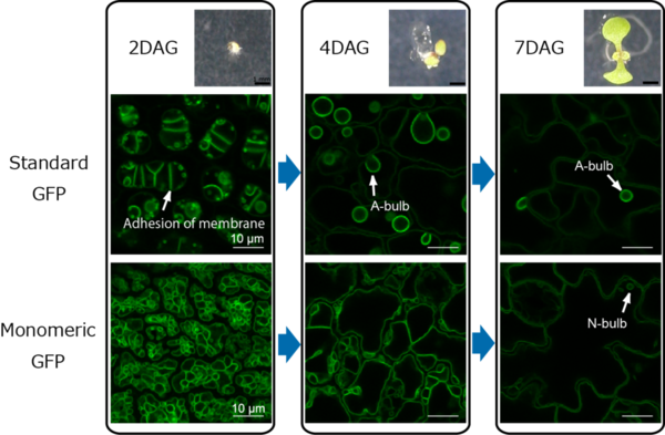 Figure 4. The image of cotyledons (i.e., seed leaves) observed by a confocal laser scanning microscope. DAG means "day after germination". A-bulb and N-bulb show an artificial bulb and a natural bulb, respectively. (Figure: by courtesy of Dr. Segami)
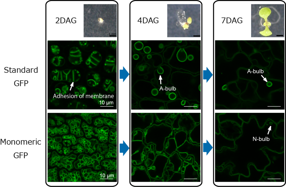
"As I expected,"
standard GFPs formed dimers and facilitated the adhesion of membranes created a strong intensity of fluorescent bulb structures, whereas monomeric GFPs did not. In addition, because a few spherical structures were observed but had different features from the creation by standard GFPs, Dr. Segami also revealed that these bulb structures certainly exist as the native intravacuolar spherical structures (Figure 4).
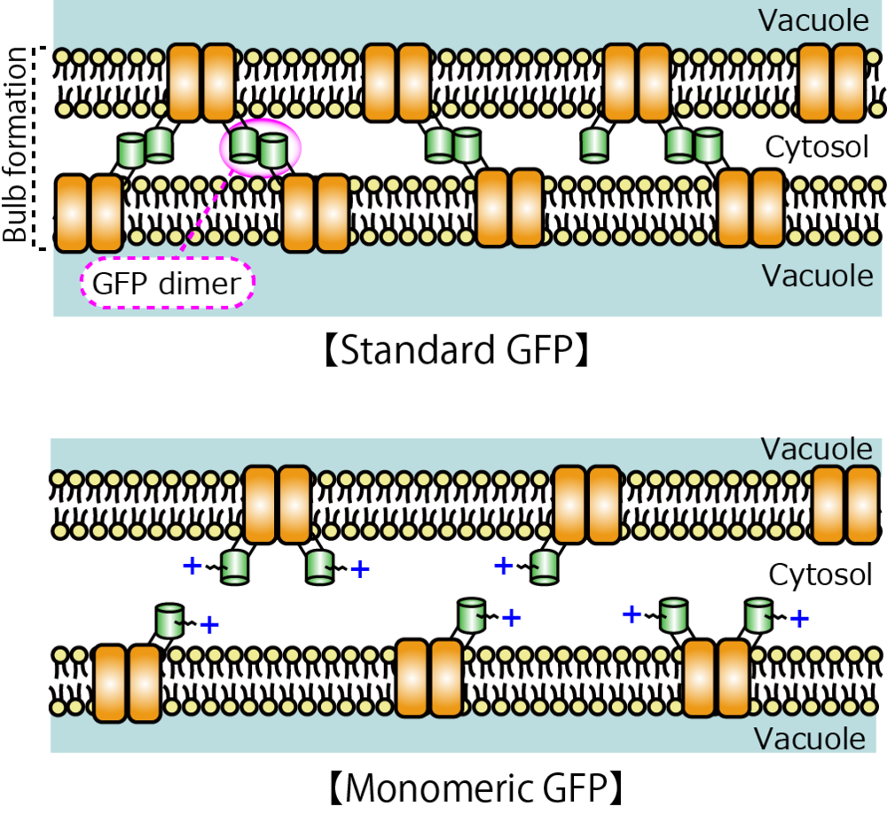
Figure 5. The schematic diagram of bulb formation by adhesion of vacuole membranes. (Figure: by courtesy of Dr. Segami)
Dr. Segami and his co-workers found and overcome the artificial phenomena by GFPs (Figure 5). As a result, they concluded the accurate features of H+-PPase that exists in young tissues or tissues with a high sugar content as well as large number of vacuoles coexisting in such environments.
Scientists who study visualized proteins across the world, regardless of biological species, should have knowledge of the properties of GFPs to increase the reliability of the use. In fact, during basic research in the field of life sciences, Dr. Segami's monomeric GFP linked with H+-PPase is considered as a vacuolar membrane marker that can trace the vacuoles naturally and clearly, and he has been flooded with a number of queries from all over the world.
*******
One of the conditions for errorless observation is the quality of the markers. Besides GFPs, the artificial interplay between target molecules with other fluorescent proteins should also not be neglected.
"The result was guided because I have observed the target molecules carefully. If we check carefully at each experimental stage, we may come across an unexpected discovery."
Dr. Segami's methodology can be applied to other fluorescent proteins.
Increasing the reliability of the use of markers
―accomplishments from basic research field surely advance science and technology.
(Ayako Umemura)
Researchers featured in this article
Dr. Masayoshi Maeshima【Professor, Graduate School of Bioagricultural Sciences, Nagoya University】

Dr. Masayoshi Maeshima was born in Shizuoka-Prefecture (current Hamamatsu-City) in 1954. He obtained a doctorate degree in agriculture from Nagoya University in 1981. In 1984, he worked as a postdoctoral researcher at the University of California and as an Assistant at the School of Agricultural Sciences, Nagoya University. In 1988, he moved to the Institute of Low Temperature Science Hokkaido University as an assistant and became an Associate Professor at the Institute. In 1994, he began his career as an Associate Professor at the School of Agricultural Sciences, Nagoya University, and between 1996 and 1998 he also conducted research at the National Institute for Basic Biology as a Visiting Associate Professor. In 1997, because of the university's structural reform, he became an Associate Professor at the Graduate School of Bioagricultural Sciences, Nagoya University and since 2001, he has remained in in the present post.
Since 2011, Dr. Maeshima has become a Trustee of the Graduate School of Bioagricultural Sciences, Nagoya University. In 2012, he became the Dean of the School of Agricultural Sciences, and the Dean of Graduate School of Bioagricultural Sciences, Nagoya University.
***
"I enjoyed fishing in rivers as a child" said Dr. Maeshima. Recently, he has been unable this because of his work schedule; although, he has enjoyed listening to music in his car.
Getting refreshed in a relaxed space, Dr. Maeshima leads researchers with a strong will, observing any kind of events carefully. I expect he will keep supervising them to discover interesting research results and finding out the real value of the study on the way to their own research purposes (by AU)
Dr. Shoji Segami【Postdoctoral researcher, Graduate School of Bioagricultural Sciences, Nagoya University】

In 2004, Dr. Shoji Segami graduated from the School of Agricultural Sciences, Nagoya University, and completed his Masters course at the Graduate School of Bioagricultural Sciences, Nagoya University. He obtained his doctorate degree from Nagoya University in 2011. Since April 2011, he has worked at his current position.
***
"I've been interested in plants since I was in elementary school" said Dr. Segami. He likes any kind of plants with trees and weeds including. His most favorite plant is maple because it is beautiful in all seasons. In addition, he likes to grow insectivores as he considers them as "weird plants."
Dr. Segami observes things carefully in both microscopic and macroscopic views in his personal life as well as during his research. I am looking forward to his future research achievements (by AU)
Links
- Laboratory HP http://celld.agr.nagoya-u.ac.jp/index-e.html
- Link to this article (from a search-result)
http://www.plantcell.org/search?author1=&fulltext=&pubdate_year=2014&volume=26&firstpage=3416&submit=yes
Shoji Segami, Sachi Makino, Ai Miyake, Mariko Asaoka, and Masayoshi Maeshima
Dynamics of vacuoles and H+-pyrophosphatase visualized by monomeric green fluorescent protein in Arabidopsis: artifactual bulbs and native intravacuolar spherical structures.
The Plant Cell 2014 26: 3416-3434.
(First published on August 12, 2014; doi:10.1105/tpc.114.127571)
- Link to related article (from a search-result)
http://www.jbc.org/content/264/33/20068
Maeshima M and Yoshida S
Purification and properties of vacuolar membrane proton-translocating inorganic pyrophosphatase from mung bean.
The Journal of Biological Chemistry 1989 264:20068-73.